Sorry, you need to enable JavaScript to visit this website.
Skip to main content

MECHANISM OF DISEASE

IgA nephropathy (IgAN) is characterized by glomerular deposition of immune complexes containing galactose-deficient IgA1 that lead to kidney damage1–5

There are 4 processes or ‘Hits’ involved in the pathogenesis of IgAN:6,7

Pathogenesis of IgA nephropathy diagram

Image adapted from Suzuki H, et al. 20116

This formation of pathogenic IgA1-containing immune complexes triggers:1–3

IgA nephropathy disease sequence diagram

Different stages of pathology in IgA nephropathy7

Stage 1 histopathologic image in IgA nephropathy
1. IgAN at an early stage, with minimal mesangial expansion and preserved glomerular capillary tufts (that is, a network of capillaries) architecture. Periodic acid-silver methanamine, haematoxylin and eosin counterstain, 360× magnification
Stage 2 histopathologic image in IgA nephropathy
2. Small segmental sclerosis (25%) with capillary collapse (thin arrow) and capsular adhesion (thick arrow). Periodic acid-silver methanamine, haematoxylin and eosin counterstain, 360× magnification
Stage 3 histopathologic image in IgA nephropathy
3. Significant segmental sclerosis (50% of the glomerular area) with capillary collapse and consolidation (*), hyalinosis (thin arrow) and capsular adhesion (thick arrow). Periodic acid-silver methanamine, haematoxylin and eosin counterstain, 360× magnification
Stage 4 histopathologic image in IgA nephropathy
4. Advanced glomerulosclerosis or glomerular obsolescence (>75%), with associated tubular atrophy and interstitial fibrosis. Periodic acid-silver methanamine, haematoxylin and eosin counterstain, 180× magnification
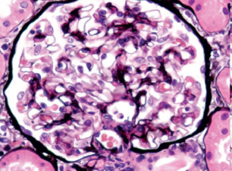
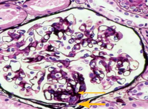
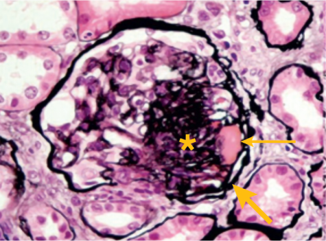
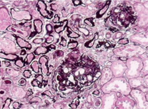
Image adapted from Lai KN, et al. 20167

DAMAGE DUE TO IgAN OCCURS THROUGH VARIOUS PATHOLOGIC PROCESSES

FIND OUT MORE